Figure 1.

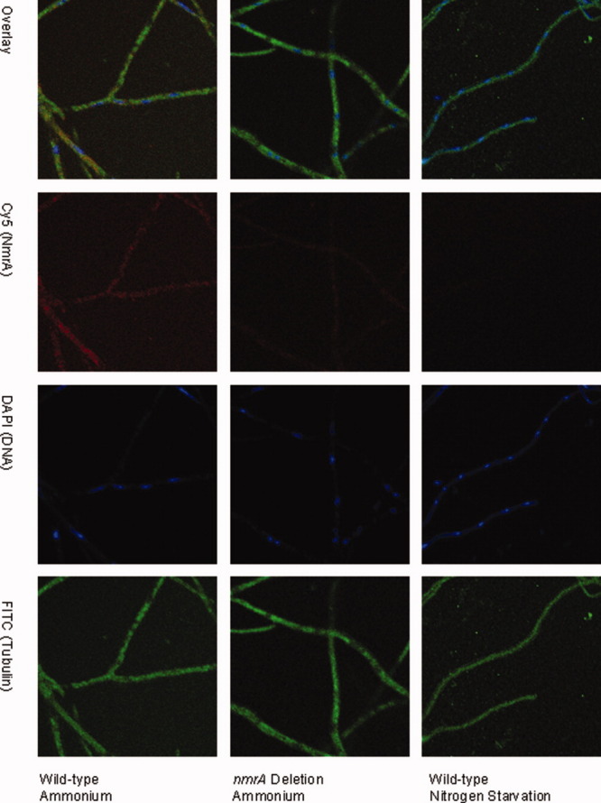
Confocal microscopy analysis of NmrA levels in wild type and nmrA deletion strains. wild type and nmrA deletion strains of A. nidulans were grown on cover slips on minimal medium containing 10 mM ammonium, 0.05% w/v glucose and 0.5% w/v quinic acid and for nitrogen starvation conditions transferred to liquid nitrogen free minimal medium. Mycelia were subsequently probed for the presence of NmrA by confocal fluorescence microscopy. Immunostaining was carried out as described in Methods with NmrA staining red, tubulin green (to delineate the boundaries of the hyphae), and DNA cyan (to define the boundaries of the nuclei). The top row shows all three images overlaid, the second row shows the fluorescence due to the presence of NmrA, the third row shows the fluorescence due to DNA staining in the nucleus and the bottom row shows the fluorescence due to staining tubulin.
