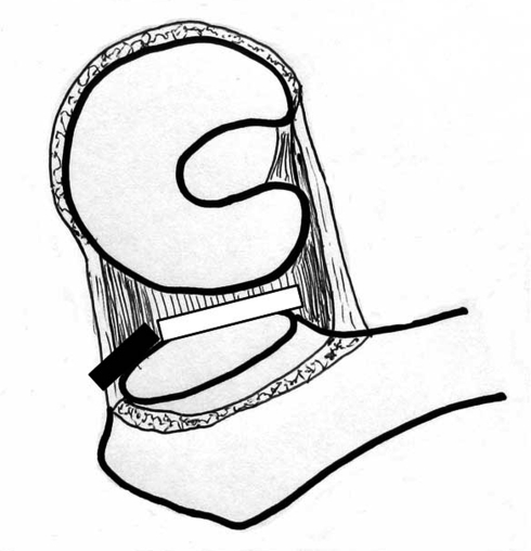
Fig. 1.
The schematic illustration demonstrates the release of the superior capsule (black bar), which was attached between the ilium and the trochanteric fossa, and the release of the posterior capsule (white bar), which was attached between the posterior rim of the acetabulum and posterior surface of the femoral neck. (This illustration was presented as Fig. 1 in Poster No. 2039 at the 55th Annual Meeting of the Orthopaedic Research Society, February 22–25, 2009 in Las Vegas, Nevada.)

