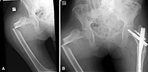
Fig. 4A–B.
Radiographs of the right hip and pelvis, respectively, show (A) a low-energy subtrochanteric fracture on the right and (B) a previous left subtrochanteric fracture in the same patient. The bilateral subtrochanteric insufficiency fracture pattern with the transverse fracture line on the tension side of the femur and lateral cortical thickening adjacent to the fracture can be seen.

