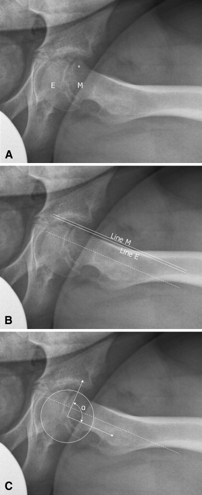
Fig. 1A–C.
Frog-leg lateral radiographs of the hip show posterior tilt and translation of the epiphysis (E) with anterior prominence (*) of the proximal metaphysis (M). (A) Left hip of Patient 2. (B) Epiphyseal-metaphyseal offset of left hip of Patient 2. (C) Alpha angle of left hip of Patient 2. Dashed line = line along center of femoral neck; Line E = line along anterior epiphysis parallel to femoral neck; Line M = line along anterior metaphysis parallel to femoral neck; α = alpha angle.

