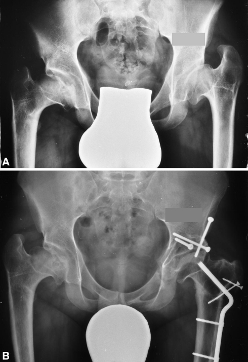
Fig. 5A–B.
(A) This AP pelvic radiograph shows bilateral sequelae of Perthes disease. A high acetabular index, mushroom-shaped femoral head, short neck and high-riding greater trochanter are apparent on the left side, although the joint space remains fairly congruent. (B) The AP radiograph 8 years after combined PAO and valgus ITO with distal advancement of the greater trochanter shows that congruency was maintained and that the acetabular index was normalized.

