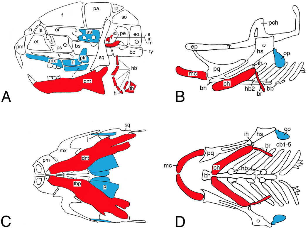
Figure 1.
Pharyngeal arch morphology in mouse and zebrafish. Lateral (A, B) and ventral (C, D) views of the craniofacial skeletons of embryonic day (E)18.5 (mouse) and 5 days postfertilization larvae (dpf) (fish). Within the mandibular and hyoid arch skeletons, Edn1 signaling is required for distal (ventral) arch cartilage and bone (red) and inhibits formation of more proximal (dorsal) cartilage and bone (blue). as, alisphenoid; bb, basibranchial; bh, basihyal; bo, basioccipital; br, branchiostegal ray; bs, basisphenoid; cb, ceratobranchial; ch, ceratohyal; dnt, dentary; eo, exoccipital; ep, ethmoid plate; et, ethmoid; f, frontal; h, hyoid; hb, hypobranchial; hs, hyosymplectic; ih, interhyal; in, incus; ip, interparietal; j, jugal; la, lacrimal; m, malleus; mc, Meckel’s; mx, maxilla; n, nasal; op, opercle; or, orbital; p, palatine; pa, parietal; pch, prechordal; pe, petrosal; pl, palatine; pm, premaxilla; pq, palatoquadrate; ps, presphenoid; ptr, pterygoid; s, stapes; so, supraoccipital; sq, squamosal; tbp, trabecular basal plate; th, thyroid; tr, trabecula; ty, tympanic ring; v, vomer.

