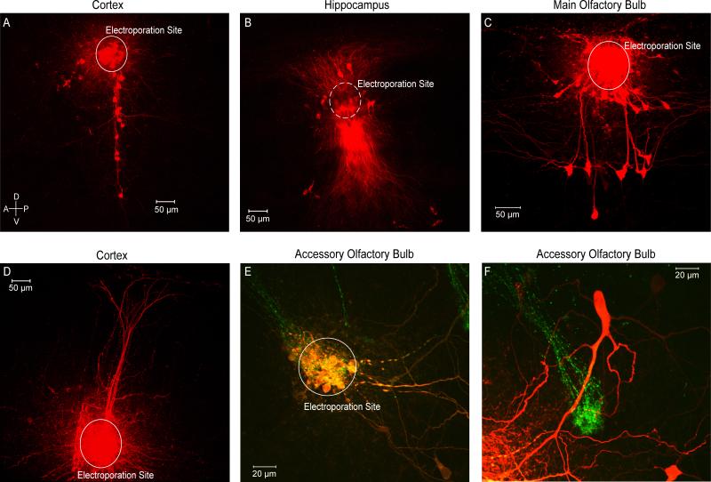
Figure 1. Electroporation of Various Circuits.
A) Coronal slice containing barrel cortex was electroporated in layer 2/3 labeling the processes of several neurons in other cortical layers. B) Coronal slice containing hippocampus was electroporated in area CA1 labeling multiple pyramidal cells and their projections. C) Sagital slice containing the main olfactory bulb. A single glomerulus was electroporated labeling periglomerular, external tufted, and mitral cells all innervating the electroporated glomerulus. D) Coronal slice containing somatosensory cortex was electroporated in layer 5 labeling pyramidal cells and their projections towards layer 1. E) Sagittal slice containing the accessory olfactory bulb in a GFP-transgenic mouse line. A GFP-positive glomerulus was targeted and electroporated. F) A mitral cell labeled by electroporating the glomerulus in E shown beside a second GFP-positive glomerulus.

