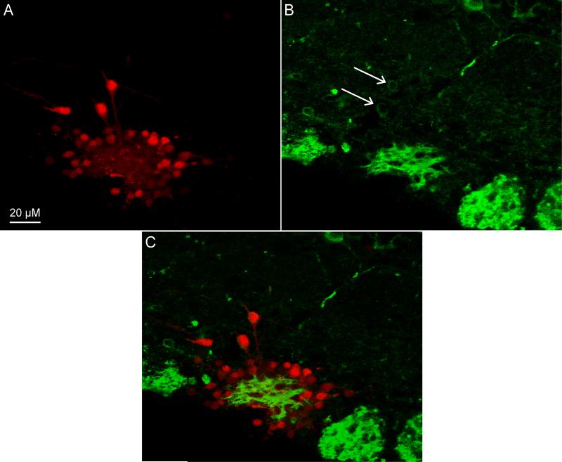
Figure 3. Electroporation-Coupled Immunohistochemistry.
A) Confocal images stack of a coronal slice containing the main olfactory bulb in which one glomerulus was electroporated labeling several periglomerular and external tufted cells. B) Confocal image stack of the same slice in A which was also stained for Kv. 1.2 potassium channels. Arrows indicated two nearby Kv. 1.2-positive cells. C) Overlay of A & B illustrating the expression of Kv. 1.2 potassium channels by these two external tufted cells which were also labeled via electroporation.

