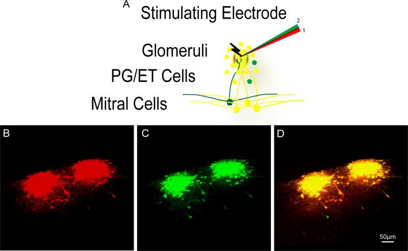
Figure 5. Completeness of Electroporation Protocol.
A) Schematic illustrating the experimental design using a theta glass electrode, each chamber filled with a differently colored dye. A given glomerulus was electroporated first with the red chamber and second with the green chamber. B) Confocal image stack of the red channel from two adjacent glomeruli which had been electroporated labeling many periglomerular, external tufted, and mitral cells. C) Confocal image stack of the green channel from the same two glomeruli in panel B. D) Overlap of both channels to observe the number of cells labeled only after the second electroporation protocol was given (green cells) and those labeled by both (yellow).

