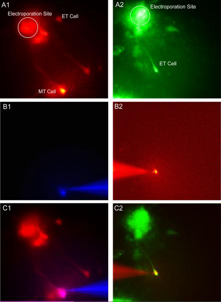
Figure 6. Targeted Whole-Cell Patch Clamp via Electroporation.
A1 & A2) Fluorescent images of two separate slices in which a single glomerulus was electroporated with Rhodamine Ruby Red (A1) and Oregon Green Bapta 488 (A2) labeling external tufted and mitral cells. B1 & B2) Fluorescence enabled targeted whole-cell patch clamping and filling with a second fluorescent dye, Alexa 350 (B1) and Alexa 594 (B2). C1 & C2) Overlay of A1 vs B1 and A2 vs B2 confirm cell identify.

