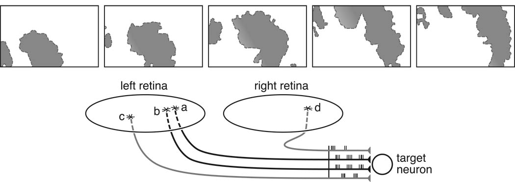
Figure 7.
Spontaneous (pre-visual) waves of activity spread across the surface of the developing retina (top), affecting each retina independently. The top five panels show the active regions (shaded) at two-second intervals from left to right. As a consequence, the bursts of action potentials by retinal ganglion cells will be more highly correlated between those axon terminals arising from adjacent locations on the retinal surface (a and b), ensuring a stabilization of their nascent synaptic contacts with target neurons, refining the precision of the retinotopic map (bottom). Much as more distant locations on the retina will have non-synchronous spontaneous activity with these two cells (c), so locations on the opposite retina should generate asynchronous discharges (d) relative to these cells, leading to the loss of binocular innervation upon single cells in the developing lateral geniculate nucleus. (Top modified from Stafford, Sher, Litke, & Feldheim, 2009).

