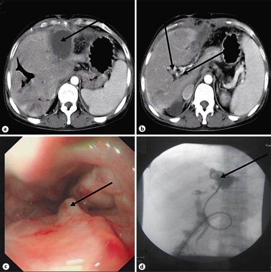
Fig. 1.
A 45-year-old female patient with CTPV and drainage for acute liver abscess. a Round hypodense lesion of liver abscess (arrow). b Dilated bile ducts with multiple stones, portal vein thrombosis and collateral circulation around the portal vein (arrows). c Esophegeal varices with F3 Ls Rc (+) E0 (arrow). d Digital subtraction angiography-guided percutaneous hepatic abscess drainage was performed within one week after the admission (arrow).

