Abstract
We present the case of a 68-year-old female patient who was diagnosed with cancer of the descending colon in July 1994 and underwent partial resection of the colon (type 2, moderately to well differentiated adenocarcinoma, se, ly1, v1, n(-)). In April 1996, she was admitted to a nearby hospital for symptoms of ileus, which improved at the hospital. However, she was referred to our hospital for melena. In blood test, Hb was 8.7 g/dl, showing anemia, and carcinoembryonic antigen level was elevated to 50.7 ng/ml. Abdominal CT and small bowel series showed only mild expansion of the small bowel, suggesting no obvious occlusion. Abdominal surgery was performed in May 1995 for repeated development of ileus symptoms and suspicion of bleeding from the small bowel. Since the findings of the abdominal surgery showed a circular tumor in the lower ileum, partial resection of the small bowel was performed. Histopathological examination showed type 3, moderately to well differentiated adnocarcinoma, se, ly2, v0, n = 1/13. The principal tumor was located within the subserosa and grew up exclusively through the muscularis propria and the submucosa, into the mucous layer. The mucosa remained slightly on the surface layer. Based on these findings, the patient was diagnosed with metastasis of descending colon cancer to the small bowel. Her prognosis was good, and neither metastasis nor redevelopment of the cancer have been confirmed to date, 11 years and 7 months since the surgery.
Key Words: Metastatic small bowel tumor, Colon cancer
Introduction
The most common primary focus of metastatic tumor of the small intestine is lung cancer, followed by breast cancer and gastric cancer. The incidence of metastasis from colon cancer is rare [1], but the majority of cases involve invasive/disseminated metastasis and there have been a few reports of hematogenous metastasis to the vascular system. We experienced a case of suspected hematogenous metastasis to the small intestine from descending colon cancer.
Case Report
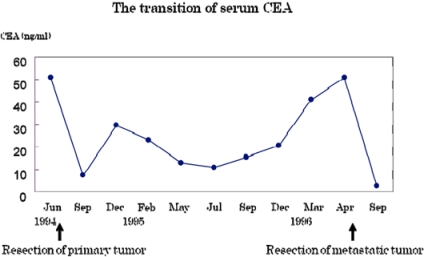
A 68-year-old female presented with the chief complaint of melena. Her medical history were hypertension (from the age of 55 years) and femoral neck fracture (at 60 years). A partial colonic resection was performed for the diagnosis of descending colon cancer in July 1994 (type 2, 45 × 35 mm, moderately to well differentiated adenocarcinoma, se, ly1, v1, n(-), H(-), P(-), M(-)). Ileus had occurred several times since October 1995 and had been alleviated by conservative therapy. She was referred to a local physician in April 1996. The symptom subsided but melena occurred. She was referred to our hospital and hospitalized for detailed examination and treatment. Status at admission was: body temperature 36.7°C, blood pressure 135/72 mm Hg, pulse rate 79/min, regular pulse. No abnormal findings were observed in the abdomen. Blood test on admission indicated Hb 8.7 g/dl, i.e. anemia. Carcinoembryonic antigen (CEA) level was elevated to 50.7 ng/ml. According to the follow-up at the outpatient department, CEA level remained high, 52 ng/ml, before the surgery for the descending colon cancer. Although the CEA level decreased after surgery, it increased slightly and ranged between 10 and 30 ng/ml (fig. 1). Abdominal X-rays revealed gas masses throughout the entire small intestine without niveau.
Fig. 1.
The transition of serum CEA.
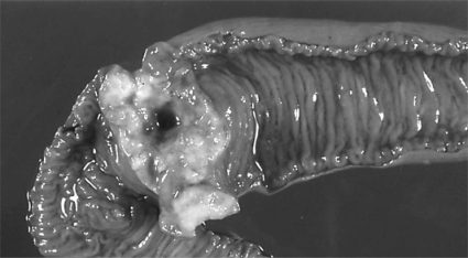
Abdominal CT revealed mild dilation of the small intestine but no findings that suggested obvious metastasis or relapse of cancer. Gastroscopy and colonoscopy showed no abnormal findings. Small bowel series revealed mild dilation throughout the whole of the small intestine but no findings that indicated obvious stenosis or occlusion. Based on the above-mentioned findings, the patient was diagnosed as having repeated ileus and possible small intestine hemorrhage, and laparotomy was performed in May 1996. Intraoperative findings demonstrated neither ascites nor peritoneal metastasis. The lesion encircled the lower ileum, where mild contraction was observed in the serosa and the mesenterium, but there was no evidence of exposed tumor. A partial small bowel resection including the tumor area was performed. Fresh specimen revealed a type 3-like circumferential tumor in the ileum, 30 × 50 mm in size (fig. 2). Mild contraction was found in the serosal surface, but there was no evidence of exposed tumor. Pathohistological examination revealed that the main lesion of the tumor was located within the subserosa and grew up exclusively through the muscularis propria and the submucosa, into the mucous layer, which was similar to the histopathologic image of the descending colon cancer isolated in 1994 (fig. 3). In addition, there were no operative findings to indicate the presence of invasive/disseminated metastasis. Therefore the patient was diagnosed as having hematogenous metastasis to the small intestine. She was discharged in excellent condition. At present, 11 years and 7 months after the surgery, neither relapse nor metastasis have been observed.
Fig. 2.
Macroscopic findings of the mucosal side of the resected small intenstine.
Fig. 3.
Histological appearance of the primary descending colon cancer (a) and metastatic tumor (b). Note the similar histological features.
Discussion
The incidence of metastatic tumors in the small intestine is relatively rare, and 2.8-8.2% have been identified in autopsy cases [2,3,4,5,6]. The routes of metastasis to the small intestine include hematogenous metastasis (a tumor grows within the intestinal wall and spreads via hematogenous or lymphatic routes), peritoneal metastasis (a tumor, which disseminates and invades into the serosa and the mesenterium, continuously increases within the intestinal wall), and intestinal metastasis (tumor cells, which are liberated/drop out into the intestinal tract, are implanted into the intestinal mucosa and grow). On the other hand, the major route of metastasis of colon cancer to the small intestine is disseminated metastasis associated with peritonitis carcinomatosa [3].
The reported number of suspected cases of hematogenous metastasis of colon cancer, like our case, was only 6 from 1983 to 2007 in Japan. We reviewed a total of 7 patients, including the above-mentioned 6 patients and our patient (table 1) [1, 7,8,9,10,11]. The ages of the patients, including 4 males and 3 females, ranged from 60 to 80 years, with a mean of 69.0 years. Therefore no sex difference was observed. Their main symptoms included obstructive symptoms, such as abdominal bloating, vomiting, and constipation, and bleeding symptoms, such as occult blood and melena caused by bleeding from tumors. In addition to tumor occlusion and hemorrhage, perforation and palpable abdominal mass are generally seen [6]. The most common primary site among the cases reviewed was the sigmoid colon in 3 patients, and the ascending colon, the transverse colon, the descending colon, and the rectum in 1 patient each. The histopathological diagnosis indicated that the number of moderately differentiated adenocarcinomas, low to moderately differentiated adenocarcinomas, and moderately to well differentiated adenocarcinomas were 5, 1, and 1, respectively, and that there were no remarkable profiles. The tumor progression was high and the depth of the tumor invasion was more than the subserosal layer in all of the patients. The hematogenous and lymphatic invasion level, ly1, v1 and greater, was observed in all of the patients. With the exception of 1 patient whose metastasis site was both the jejunum and the ileum, the metastasis site of the remaining patients was the ileum in 5 patients and the jejunum in 1 patient. Thus, the ileum was the most common metastasis site. One patient in the review had 3 metastases in total, including 1 in the jejunum and 2 in the ileum, and 1 patient had 2 metastases in the ileum. The remaining patients had only 1 metastatic focus.
Table 1.
Reported cases of metastatic small bowel tumor from colon cancer with extensive hematogenous or lymphogenous spread in Japan
| First author | Age/sex | Chief complaint | Locationa | Histology | Durationb | Locationc | Prognosis |
|---|---|---|---|---|---|---|---|
| Yamamoto (1997) [8] | 76/M | abdominal distention | S/C | mod, ss, ly3, vl, nl(+) | 9 years | ileum | alive (13 months), no recurrence |
| Niwa(2003) [9] | 69/F | vomiting | T/C | mod, ss, ly2, v2, n(−) | 3 years | jejunum | alive (6 months), no recurrence |
| Ishida(2003) [10] | 80/F | constipation | A/C | poor-mod | same time | ileum | death (1 month) |
| Kuroda(2005) [1] | 62/M | general fatigue | S/C | mod, ss, lyl, v3 | same time | jejunum and ileum | alive (31 months), no recurrence |
| Takeshita(2006) [11] | 60/M | constipation | rectum | mod, ss, lyl, vl, n3(+) | 2.5 years | ileum | alive (30 months) |
| Tsujimura (2007) [7] | 68/M | none | S/C | mod, ss, ly2, v2, nl(+) | 2 years | ileum | alive (18 months) |
| Our case | 68/F | melena | D/C | mod-well, se, lyl, vl, n(−) | 1.6 years | ileum | alive (134 months), no recurrence |
Location of primary lesion.
Duration before detection of metastatic tumor.
Location of metastatic lesion.
In terms of time to diagnosis, synchronous metastasis was observed in 2 patients, and metachronous metastasis took from 1 year and 8 months to 9 years, with a mean of 3 years and 7 months after surgery. One patient had metastasis to the lung and the liver and all of the remaining patients had metastasis to the small intestine alone. The majority had a solitary metastasis.
The most common method of clinical diagnosis of metastatic carcinoma of the small intestine is a small bowel series. The typical findings are that (1) it has a submucosal tumor with a clear-cut margin and central depression called the bull's eye sign, and that (2) it exhibits fold convergency transverse to the longitudinal axis of the bowel lumen (transverse stretch) [6]. However, in this review there were no cases in whom a preoperative diagnosis was established. They were found during surgery for intestinal obstruction in 3 patients, during surgery for the primary focus in 2 patients, and during surgery for anastomotic recurrence of the primary focus in 1 patient. Our case underwent surgical treatment for repeated intestinal obstruction and small intestinal bleeding, but there was no remarkable abnormality, so that a preoperative definitive diagnosis was not established. The surgical procedures conducted in this review were ileocecal resection in 3 patients, partial small bowel resection in 3 patients, and right hemicolectomy in 1 patient. All of the patients underwent tumor resection containing the primary focus.
The histopathological findings in the cases of hematogenous metastasis to the small intestine indicated distant metastasis to the submucosa and/or muscularis propria and temporal increase of the primary focus in the mucosal and serosal sides. Therefore, unlike tumors that invade directly and metastasize to the small intestine, a primary focus close to the serosa may develop into an extra-gastrointestinal tumor or submucosal tumor, whereas a primary metastasis close to the mucosal side is likely to form an ulcer but may have partially retained morphology of a submucosal tumor [8]. All 10 of the primary foci studied in this review included 5 submucosal tumor-like lesions, 2 type 2-like lesions, 1 type 3-like lesion, 1 type 1-like lesion, and 1 cerebriform lesion.
In general, it has been reported that the prognosis of metastasis to the small intestine is poor. Metastatic tumors are normally found as a result of the presence of remarkable symptoms, such as gastrointestinal bleeding, intestinal obstruction, and intestinal perforation, and these severe findings may lead to the diagnosis [2, 5, 6]. However, in this review, 4 patients had neither relapse nor metastasis at 6 months, 1 year and 1 month, 2 years and 7 months, and 11 years and 2 months, respectively, and 2 patients survived for 1 year and 6 months and 2 years and 6 months, except for one death that occurred 1 month after surgery. Accordingly, the long-term prognosis can be expected to improve by resection of the primary focus.
All cases in this review, except for the cases in whom the metastatic tumor was found accidentally at the time of operation, had intestinal occlusion. Postoperative intestinal occlusion is very familiar to surgeons. Tanaka et al. [12] reported that the percentage of adhesive intestinal obstruction, ileus caused by peritonitis carcinomatosa, and occlusive ileus caused by tumor was 60.9, 18.5, and 10.7%, respectively, among cases of obturation ileus. When encountering postoperative intestinal occlusion, which results from a malignant tumor, the possibility of metastasis to the small intestine as well as adhesive intestinal obstruction should be considered.
Footnotes
This is an Open Access article licensed under the terms of the Creative Commons Attribution-NonCommercial-NoDerivs 3.0 License (www.karger.com/OA-license), applicable to the online version of the article only. Distribution for non-commercial purposes only.
References
- 1.Kuroda M, Tanaka Y, Usami K, Yoshitani S, Kita I, Takashima S. A case of multiple sigmoid colon cancer which metastasized to the small intestine. J Jpn Surg Assoc. 2005;66:1119–1124. [Google Scholar]
- 2.Masaoka K, Nakamura T. Metastatic neoplasms of the bowel. Nippon Rinsho. 1994;(suppl 6):612–615. [PubMed] [Google Scholar]
- 3.DeCastro CA, Dockerty MB, Mayo CW. Metastatic tumors of the small intestines. Surg Gynecol Obstet. 1957;105:159–165. [PubMed] [Google Scholar]
- 4.Farmer RG, Hawk WA. Metastatic tumors of the small bowel. Gastroenterology. 1964;47:496–504. [PubMed] [Google Scholar]
- 5.Routh A, Hickman BT. Metastatic tumors of the small intestine: case report and review of literature. J Miss State Med Assoc. 1984;25:235–236. [PubMed] [Google Scholar]
- 6.Ushio K, Ishikawa T, Miyagawa K. X-ray diagnosis of metastatic small intestinal tumor. I to Cho. 1992;27:793–804. [Google Scholar]
- 7.Tsujimura T, Toyokawa A, Wakahara T, Mukubou H, Hamabe Y. A case of solitary metastasis of sigmoid colon cancer to the small intestine. Jpn J Gastroenterol Surg. 2007;40:141–145. [Google Scholar]
- 8.Yamamoto M, Karasawa G, Hirata K, Konishi Y, Takada Y, Yokoyama S. A case of strongly-suspected hematogenous metastasis after surgery for sigmoid colon cancer. Hokkaido J Surg. 1997;42:199–200. [Google Scholar]
- 9.Niwa H, Morikane K, Naka S, Morikane K, Nka H, Yasuhara H. Recurrent tumor of the small intestine after transverse colon cancer surgery. Shujyutsu. 2003;57:527–531. [Google Scholar]
- 10.Ishida T, Kato T, Ito Y, Hukuhara R, Yamazaki M. A case of possible synchronous lymph node metastasis of ascending colon cancer to the small intestine. Mie-Igaku. 2003;46:81–84. [Google Scholar]
- 11.Takeshita H, Tsuji T, Sawai T, Takasaki H, Deguchi M, Yasutake T, Nagayasu T. A case of a metastatic jejunal tumor that developed intestinal occlusion 2.5 years after surgery for rectal cancer. J Jpn Surg Assoc. 2006;67:640–644. [Google Scholar]
- 12.Tanaka N, Onda M, Takasaki H, Yoshimura K, Yokoyama S, Sasabe H, Nagashima Y. Cause and pathophysiology of ileus. Shokaki Geka. 1996;19:1787–1792. [Google Scholar]


