
Figure 4.
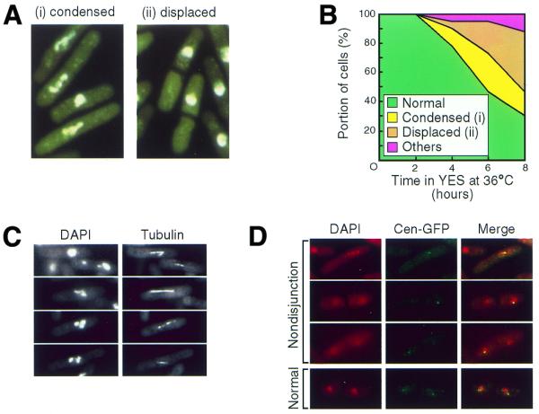
Defective chromosome segregation in the dhp1-1 mutant. (A) Nuclear structures of the dhp1-1 mutant at 36°C. dhp1-1 (MP102) cells grown at 36°C for 6 h were stained with DAPI to visualize the nucleus. Typical nuclear morphologies of defective chromosome segregation are indicated: (i) cells with condensed chromosomes and (ii) cells with displaced nuclei. See also (C) and (D). (B) Frequency of the dhp1-1 cells exhibiting defective chromosome segregation. The dhp1-1 cells grown at 26°C were cultured at 36°C and aliquots were fixed with 70% ethanol and stained with DAPI. At least 120 cells were analyzed at every interval. (C) Elongating mitotic spindles that failed to segregate chromosomes. The dhp1-1 cells cultured at 36°C for 6 h were subjected to immunofluorescence microscopy with the monoclonal anti-tubulin antibody YOL1/34. (D) Nondisjunction of sister chromatids in the dhp1-1 mutant. dhp1-1 cells (KP38) were cultured at 36°C for 6 h and the centromere DNA of chromosome I in the dhp1-1 mutant was visualized by using the Cen1–GFP system.