
Figure 4. Dye transfer between NRVM and hAFS cells.
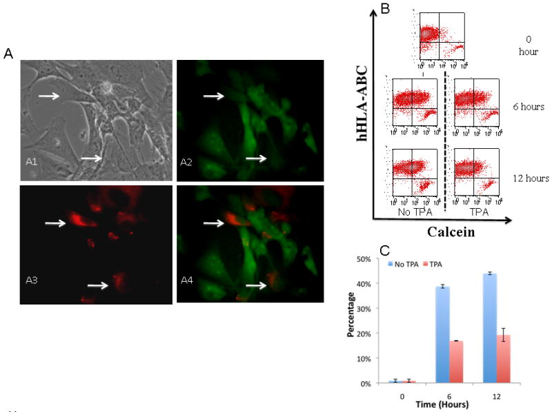
hAFS cells were cultured together with NRVM preloaded with calcein-AM. (A1-A4) Indicate that PKH-26 labeled hAFS cells (arrow) became calcein positive when cultured with NRC. (B) FACS analysis shows that the number of calcein positive human cells (anti-hHLA-ABC antibody was used to detect human derived cells) increased over time while treatment with TPA partially inhibits the transfer of calcein. (C) Quantitative summary shows calcein positive human cells increased over time while TPA partially blocked the transfer. Values represent means ± SD