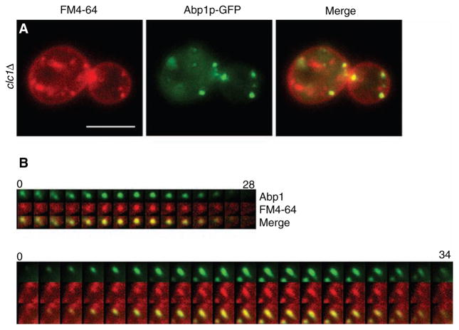
Figure 5. Actin comet tails and patches in clathrin mutants contain endocytic membrane.
(A) clc1Δ ABP1-GFP (SL5101) cells stained with 40 μM FM4-64 and incubated for 20 min before viewing using wide-field microscopy. Note patches of Abp1p associated with FM4-64 seen as yellow in the merge. (B) Two examples of time-lapse videos showing concentration of FM4-64 into de novo actin patches (top panel) and actin comet tails (bottom panel) from clc1Δ cells. Note first three frames in the top left corner of B), top panel show a residual disappearing Abp1p patch different than the de novo patch that appears in the middle of frame number 3. Scale bar = 5 μm. Time scales in (B) are in seconds.

