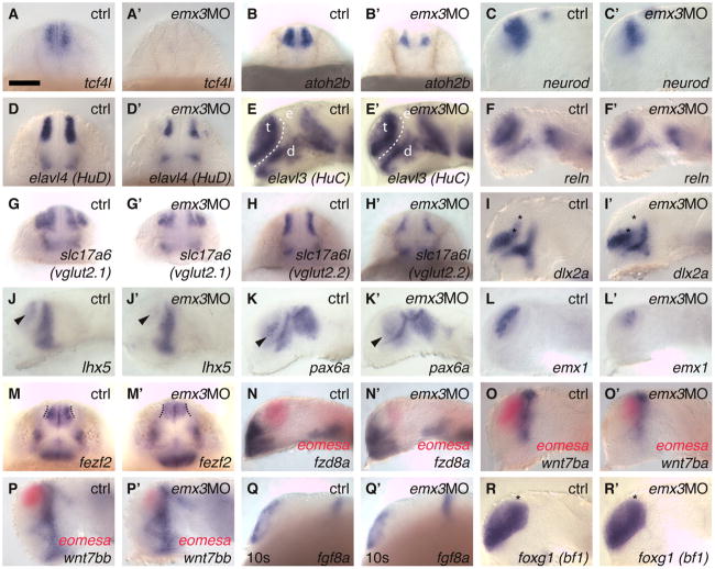
Fig. 4.
A–R: emx3 morpholino injection specifically reduces expression of dorsal telencephalic marker genes. A–D′, G–N′: Expression of tcf4l (A–A′) in the dorsal telencephalon is lost, and expression domains of atoh2b (B–B′), neurod (C–C′), elavl4 (D–D′), slc17a6 (G–G′) slc17a6l (H–H′), dlx2a (I–I′, only the domain between asterisks), lhx5 (J–J′), pax6a (K–K′), emx1 (L–L′), are reduced in emx3 morpholino antisense oligonucleotides (MO) injected embryos compared with control embryos. More broadly expressed neuronal differentiation markers elavl3 (E–E′) and reln (F–F′) show little or no apparent difference between controls and emx3MO injected embryos. fezf2 (M–M′) expression is reduced only in the more intense expression domain adjacent to the olfactory placodes (dotted lines), but unchanged in the ventricular zone. Ventral telencephalic expression domains (dlx2a, I–I′, fzd8a, N–N′; fgf8a, Q–Q′; foxg1, R–R′) are unaffected by emx3MO injections, as are diencephalic expression domains (C–K′, M–Q′). Shape and size of the telencephalon appears unaltered in emx3MO embryos compared with control embryos, as indicated for example by elavl3 (E–E′; t, telencephalon, outlined by white dotted lines; e, epiphysis; d, diencephalon), and foxg1 (R–R′) expression. Commonly used previous gene names of recently renamed genes are given in parentheses. A–B′, D–D′, G–H′, M–M′: Frontal views, dorsal to the top. C–C′, E–F′, I–L′, N–R′: Side views, dorsal to the top, rostral to the left; eyes removed except in Q–Q′. A–P′, R–R′: At 24 hr, (Q–Q′) 10-somite stage (14 hr). Scale bar = 100 μm.

