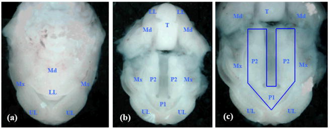
Figure 1. Photomicrographs of ventral views of the developing orofacial region of a GD-13 mouse embryo.

(a) upper and lower lips and jaws (maxilla and mandible); (b) the embryonic oral cavity; the lower half of the photo contains the roof of the oral cavity with the maxillary processes, primary palate and secondary palatal processes; the upper half contains the base/floor of the oral cavity showing the tongue and the mandible; (c) a magnified view of the roof of the oral cavity: note that the upper lip and the primary palate are completely formed, and the developing secondary palatal shelves are derived from the medial aspect of each maxillary process. The region demarcated by the blue line was excised from GD-13 embryos for extraction of total RNA. Corresponding regions were dissected from the developing orofacial region of GD-12 and GD-14 embryos. (UL) upper lip; (LL) lower lip; (Mx) maxilla; (Md) mandible; (P1) primary palate; (P2) secondary palate; (T) tongue.
