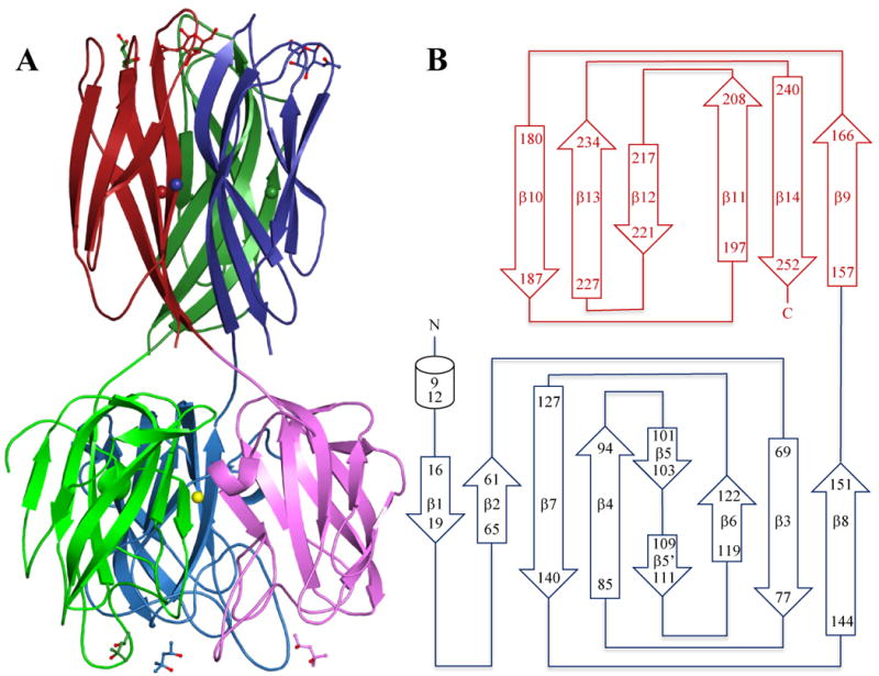
Figure 3.

A: Crystal structure of the DiscI trimer coloured by chain with the N-terminal domain in light colours and C-terminal domain in dark colours. MPD and GalNAc ligands are represented in ball and sticks. Calcium ions are represented as spheres according to chain colours and nickel ions as a yellow sphere. B: Schematic representation of DiscI topology.
