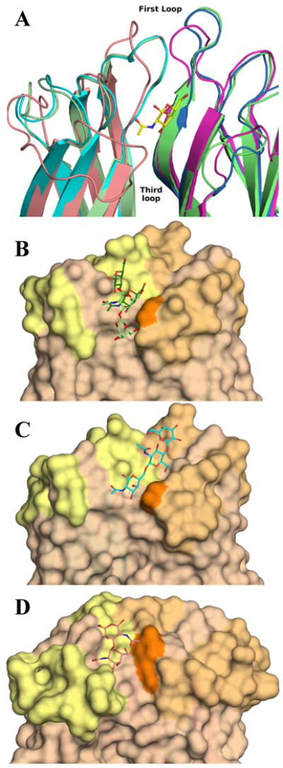
Figure 7. Binding site of H-type lectins.

A: Cartoon representation of the superimposition of DiscI (light green/green), DiscII (cyan/blue) and HPA (salmon/magenta) crystal structures. Zoom on one binding site form by two monomers coloured by chain and representation of the GalNAc moiety bound to DiscI in ball and sticks. Surface representation of the binding site of DiscI (B), DiscII (C) and HPA (D). The disaccharides, the modelled trisaccharide and Forsman antigen bound to DiscI, DiscII and HPA (PDB 2CGY) respectively are represented in ball and stick. The Galβ1-3GalNAc and GalNAcβ1-3Gal disaccharides are coloured in light and dark green respectively.
