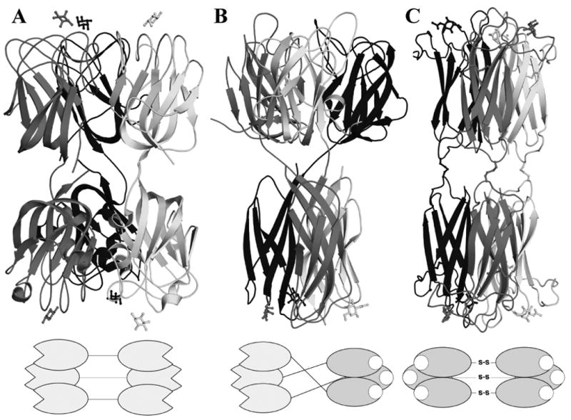
Figure 8.

Cartoon representation of the quaternary structure of the F-Lectin from striped Bass (PDB 3CQO, A), Discoidin I (B) and HPA (PDB 2CCV, C). A diagram of the domain layout is drawn beneath each protein.

Cartoon representation of the quaternary structure of the F-Lectin from striped Bass (PDB 3CQO, A), Discoidin I (B) and HPA (PDB 2CCV, C). A diagram of the domain layout is drawn beneath each protein.