
Figure 3.
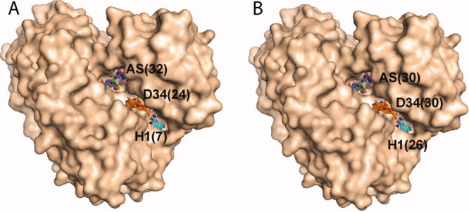
Hot spots AS, D34, and H1 in the interface of domains 3 and 4 of PMM/PGM. Numbers in parentheses show the number of probe clusters in each of the hot spots. A. PMM/PGM structure 1P5D, cocrystallized with G1P. The binding of the substrate, phosporylated at the O1 position, results in a small number of probe clusters at H1. B. PMM/PGM structure 1P5G, cocrystallized with G6P. The binding of the substrate phosporylated at the O6 position results in a much larger number of probe clusters at H1, despite the ∼20 Å distance between H1 and bound ligand.