FIGURE 2.
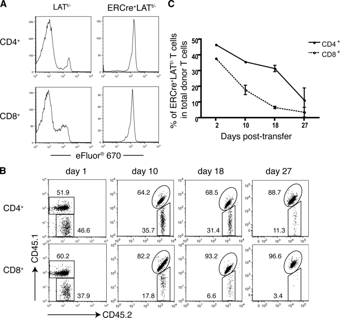
LAT deletion impairs T cell homeostatic proliferation and long term survival. A, LAT in homeostatic proliferation. Splenocytes from tamoxifen-treated LATf/− and ERCre+LATf/− littermates were labeled with Cell Proliferation Dye eFluor 670 and subsequently transferred into LAT−/− mice via tail vein injection. Seven days later, the eFluor 670 signal in CD4+ (top panel) and CD8+ (bottom panel) T cells was analyzed by flow cytometry. ERCre+LATf/− cells were gated on GFP+ cells. Data are one representative of three independent experiments. B and C, LAT in T cell survival. Splenocytes from tamoxifen-treated CD45.1+CD45.2+ ERCre+LATf/+ and CD45.2+ ERCre+LATf/− mice were mixed. The initial ratio of CD45.1+CD45.2+ to CD45.2+ CD4+GFP+ T cells was 1:1. The cell mixtures were transferred to CD45.1+ syngeneic B6 hosts. Data shown are representative of three independent experiments with three mice per experiment. B, at the indicated days following transfer, the CD45.1 versus CD45.2 expression on CD4+GFP+ (top panel) or CD8+GFP+ (bottom panel) donor T cells was analyzed by flow cytometry. The data shown are from one representative of nine mice analyzed. The numbers represent the average percentages of the gated populations. C, percentages of CD45.2+ LAT-deficient CD4+ (solid line) or CD8+ (dotted line) cells among total donor CD4+ or CD8+ T cells on the indicated days post-transfer.

