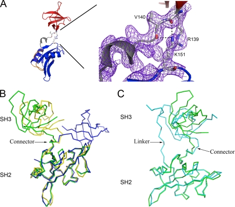
FIGURE 3.
Comparison of Src family kinase SH3-SH2 domain architecture. A, interdomain contacts in Hck32L involve numerous salt bridges (denoted by black dashes) between residues Arg-139/Val-140 (depicted as balls-and-sticks) in the SH3 and Lys-151 in the SH2 domains. These residues have well defined conformations as shown by the 2Fo − Fc electron density map (purple mesh), generated in Coot (20), and contoured at 1σ. B, comparisons of the tandem SH3-SH2 domain organizations in Hck32L (green), Fyn32 (yellow), and Lck32 (blue). C, comparison of the SH3-SH2 domain organization in Hck32L (green) to the down-regulated near full-length Hck (cyan) (PDB identifiers as indicated in Fig. 2).

