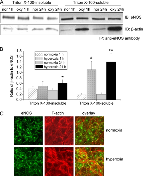
FIGURE 3.
The effects of hyperoxia on eNOS-actin association in PAEC. A and B, PAEC were exposed to 95% oxygen for 1–24 h and then Triton X-100-insoluble and soluble fractions were separated and lysed in RIPA buffer. The cell lysates from the Triton X-100-insoluble and soluble fractions were subject to co-immunoprecipitation using anti-eNOS antibody as described under “Experimental Procedures.” A is a representative blot from three separate experiments. B is a bar graph depicting the ratio of eNOS to β-actin protein in the immunoprecipitates. Results are expressed as mean ± S.D.; n = 3 experiments. *, p < 0.05 versus normoxia. C, PAEC were exposed to 95% oxygen for 24 h and then immuno-stained for eNOS (green) and F-actin (red). Images are representative of three independent experiments.

