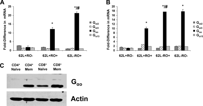
FIGURE 2.
Gαo mRNA and protein expression is highest in the most differentiated subsets of human CD4+ and CD8+ T cells. CD4+ (A) and CD8+ (B) T cells from peripheral blood were sorted into naive (CD62L+CD45RO−), TCM (CD62L+CD45RO+), and TEM (CD62L−CD45RO+; CD62L−CD45RO−) subsets as described under “Experimental Procedures.” Each subset was analyzed by semiquantitative real-time RT-PCR for mRNAs for Gαi2, Gαi3, Gαo, and Gα13. -Fold differences in the abundance of the indicated mRNAs, each normalized to that of GAPDH mRNA, between each subset of CD4+ (8) (A) or CD8+ (B) T cells were calculated for each mRNA species as described in the legend for Fig. 1. For each mRNA of interest, the lowest level of expression versus GAPDH from a single well was set at 1. Values cannot be compared between mRNA species. The data shown are the mean -fold differences ± S.E. in expression for the indicated mRNAs from assays performed in duplicate from separate donors for CD4+ and CD8+ T cells and are representative of five and three donors for CD4+ and CD8+ T cells, respectively. *, p < 0.05, when compared with mRNA in naive subsets; #, p < 0.05 when comparing mRNA content of the CD62L−CD45RO+ subset with that of the CD62L+CD45RO+ subset. C, whole-cell lysates (40 μg of protein) of purified subsets of CD4+ and CD8+ T cells, isolated as described under “Experimental Procedures,” were analyzed by Western blotting with anti-Gαo and anti-actin antibodies. Results are for naive and combined memory subsets (CD4+ Mem, CD8+ Mem) of CD4+ or CD8+ T cells. Data are from the same donor for both CD4+ and CD8+ T cell subsets and are representative of three donors.

