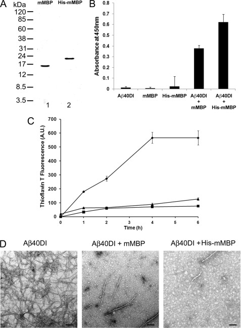
FIGURE 2.
Inhibition of Aβ fibril formation by purified mouse brain and recombinant His-tagged mouse MBP. A, MBP was either purified from mouse brain (lane 1) or recombinantly expressed in bacteria as a His-tagged protein (lane 2) and assessed by SDS-PAGE. B, interaction of Aβ40DI with mouse brain MBP (mMBP) or His-tagged mouse MBP was analyzed by solid phase binding assay. The data shown are the mean ± S.D. of triplicate determinations. C, inhibition of Aβ40DI (12.5 μm) by purified mouse brain MBP (1.56 μm) or purified His-tagged mouse MBP (1.56 μm) as assessed by thioflavin T binding and fluorescence. Aβ40DI alone, ♦; Aβ40DI + mouse MBP, ■; Aβ40DI + His-tagged mouse MBP, ▴. The data shown are the mean ± S.D. of triplicate determinations. D, TEM analysis of Aβ40DI with and without mouse brain MBP or His-tagged mouse MBP. Both forms of mouse MBP inhibit Aβ40DI fibril formation. Scale bars, 100 nm. A.U., absorbance units.

