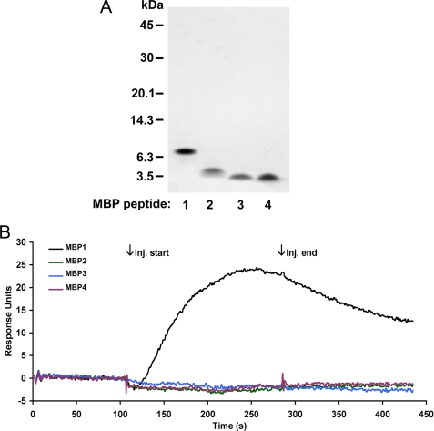
FIGURE 4.
Binding of MBP peptides to Aβ as assessed by SPR binding analysis. A, each of the MBP peptides was expressed in bacteria, purified, and assessed by SDS-PAGE. B, each of the purified MBP peptides (50 nm) was passed over immobilized Aβ40DI ligand. Representative sensorgrams were base line-corrected and plotted as overlays. Binding is identified by an increase in response during injection (Inj.) (association) followed by a gradual decrease in response (dissociation). MBP1, black line; MBP2, green line; MBP3, blue line; MBP4, red line). Only MBP1 demonstrated binding to the Aβ40DI ligand.

