FIGURE 5.
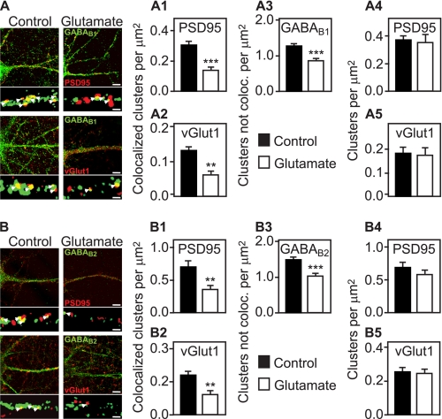
GABAB receptors associated with synapses are down-regulated by activation of glutamate receptors. Neurons were incubated in the presence or absence of 50 μm glutamate for 30 min, fixed, and subjected to double labeling immunocytochemistry using antibodies directed against GABAB1 (A, green) or GABAB2 (B, green) and antibodies against glutamatergic postsynaptic sites (PSD95, red) or glutamatergic presynaptic sites (vGlut1, red), respectively. Large images depict overviews of immunoreactivities in dendrites of lower magnification (scale bars, 5 μm), and the small images below show sections of dendrites after being processed for counting of clusters at high magnification (see “Experimental Procedures” for details; scale bars, 1 μm). In the absence of glutamate, GABAB clusters frequently colocalized (yellow, arrowheads) with either pre- or postsynaptic markers. Glutamate treatment resulted in a significant loss of GABAB1 and GABAB2 clusters associated with either PSD95 (A1 and B1) or vGlut1 (A2 and B2). GABAB receptor clusters not associated with pre- or postsynaptic marker proteins (which also include intracellular receptors) were found to be reduced to a lesser extent (A3 and B3). The number of PSD95 (A4 and B4) and vGlut1 (A5 and B5) clusters was not affected by glutamate. Mean ± S.E., n = 24 neurons derived from two independent experiments. **, p < 0.01; ***, p < 0.001, t test.

