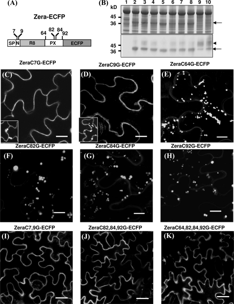
FIGURE 2.
Role of Cys residues in Zera-PB formation. A, diagram showing the positions of the cysteine residues in Zera fused to ECFP. B, total protein analysis of tobacco leaves transformed with Zera-Cys mutants fused to ECFP by SDS-PAGE/Coomassie Blue staining (top) and immunoblot using an anti-GFP antibody (bottom). Shown are untransformed tobacco protein extracts (lane 1), tobacco extracts of non-mutated Zera-ECFP (lane 2), and protein extracts of transformed tobacco with Zera Cys7 mutant (lane 3), Zera Cys9 (lane 4), Zera Cys64 (lane 5), Zera Cys82 (lane 6), Zera Cys84 (lane 7), Zera Cys92 (lane 8), Zera Cys7-Cys9 (lane 9), and Zera Cys82-Cys84-Cys92 (lane 10). The arrows indicate the electrophoretic bands of Zera-ECFP and Zera-ECFP Cys mutants, and the arrowhead shows an additional immunoreactive band. C–K, confocal images showing the fluorescence pattern of epidermal cells transformed with Zera-Cys mutants. Single Cys7 and Cys9 mutants were mainly secreted (C and D), but some cells were able to induce PBs (insets in C and D). E–H, single mutation of Cys64, Cys82, Cys84, or Cys92 had no effect on PB formation. I–K, Zera-ECFP containing multiple Cys mutations was not able to oligomerize and was secreted. Scale bars, 10 μm (C–H) or 20 μm (I–K).

