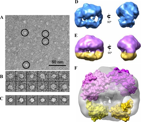
FIGURE 2.
Three-dimensional structure of BzdR using negative staining electron microscopy. A, electron microscopy field of purified His6-BzdR protein. Some representative molecules have been highlighted within circles. Scale bar, 50 nm. B, gallery of 12 selected molecules from the data set of 5,741 molecules. C, some reference-free class averages obtained using XMIPP (38). D, two views of the three-dimensional structure of BzdR reconstructed without assuming any symmetry. E, two views of the three-dimensional structure of BzdR reconstructed after imposing 2-fold symmetry. The big (violet) and the small (yellow) domains might correspond to the C-BzdR and N-BzdR domains, respectively. F, two copies of the structural models of C-BzdR (violet) and N-BzdR (yellow) can be fitted into the three-dimensional structure obtained by electron microscopy (shown as a gray transparent density), indicating that BzdR has sufficient volume to accommodate two copies of each domain. The precise orientation of the domains cannot be determined at the resolution of this three-dimensional map, and the two atomic models have been represented as a surface just to reveal the occupancy of the EM density.

