
FIGURE 3.
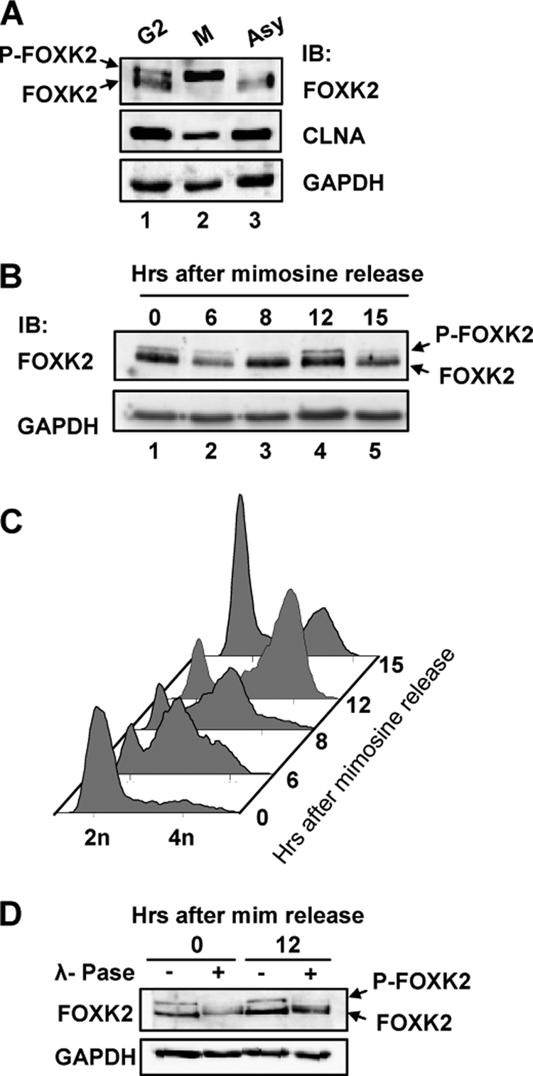
Cell cycle-dependent FOXK2 phosphorylation. A, Western blot analysis of FOXK2 during G2 and M phases. Nocodazole-synchronized mitotic U2OS cells were spilt into attached cells (G2) and mitotic cells (M) by mitotic shake off, and FOXK2 protein expression was compared with asynchronously growing cells (Asy). Protein bands corresponding to non-phosphorylated and phosphorylated FOXK2 (P-FOXK2) are indicated by arrows. Immunoblot was performed with the indicated antibodies. B and C, U2OS cells were synchronized with mimosine and released for the indicated times. The expression of endogenous FOXK2 and GAPDH were detected by immunoblot (IB) (B), and DNA content was analyzed (C). D, cell lysates from the indicated time points were treated with λ-phosphatase (λ-Pase) and probed for FOXK2 and GAPDH levels by Western blot analysis. mim, mimosine.