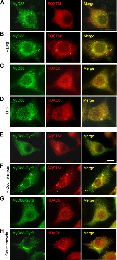
FIGURE 4.
SQSTM1 and HDAC6 are colocalized with MyD88 aggregated structures. A–D, RAW264.7 cells were left untreated (A and C) or stimulated with 1 μg/ml LPS for 1 h (B and D). Cells were then fixed and stained with anti-MyD88 rabbit polyclonal antibody and Alexa488-conjugated anti-rabbit IgG antibody and then with anti-SQSTM1 mouse monoclonal antibody (A and B) or anti-HDAC6 mouse monoclonal antibody (C and D) and Alexa564-conjugated anti-mouse IgG antibody. Scale bar, 10 μm. E–H, RAW264.7 cells stably expressing FLAG-MyD88-GyrB were left untreated (E and G) or stimulated with 1 μm coumermycin for 30 min (F and H). Cells were then fixed and stained with anti-FLAG mouse monoclonal antibody and Alexa488-conjugated anti-mouse IgG antibody and then with anti-SQSTM1 rabbit polyclonal antibody (E and F) or anti-HDAC6 rabbit polyclonal antibody (G and H) and Alexa564-conjugated anti-rabbit IgG antibody. Scale bar, 10 μm.

