FIGURE 8.
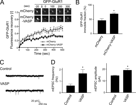
VASP enhances the retention of GluR1 in spines to potentiate synaptic strength. A, neurons were transfected with GFP-GluR1 and either mCherry or mCherry-VASP at day 6 in culture and subjected to FRAP at day 10. Prebleach and subsequent recovery images of GFP-GluR1 are shown (upper panels). The bleached point is indicated (arrow). Bar, 1 μm. To calculate the fluorescent recovery, the normalized intensity was divided by the extent of bleaching (graph). B, quantification of the immobile fraction of GFP-GluR1 clusters in cells co-transfected with mCherry or mCherry-VASP is shown. Error bars represent S.E. for 19–31 spines. *, p < 0.005). C, shown are representative traces of mEPSCs recorded from a GFP-VASP expressing neuron and an untransfected neighboring cell (Control). D, quantification of the mEPSC frequency and amplitude in GFP-VASP expressing and control untransfected neurons is shown. Error bars represent S.E. for 15-paired neurons from 11 separate experiments (*, p < 0.01).

