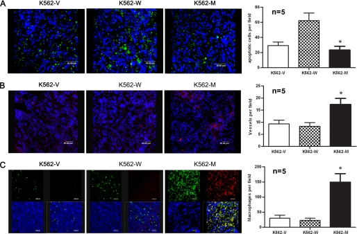
FIGURE 6.
Tumor tissue analysis. Sections were obtained from nude mouse tumor tissues formed by K562-V, K562-W, and K562-M cells on day 21 for confocal analysis. A, apoptotic cells in paraffin-embedded tumor sections were stained using the DeadEndTM fluorometric TUNEL system. B, vessels in frozen tumor sections were stained with PE-conjugated anti-mouse CD31 antibody. C, macrophages in tumor sections were stained with FITC-conjugated anti-mouse F4/80 and Alexa Fluor-conjugated anti-mouse CD206 antibodies. Quantitative analysis of apoptotic cells, vessel density, and macrophages was performed by counting 10 random high power fields from five different tumor samples within each group, and the results are shown on the right. *, p < 0.05 by Student's t test.

