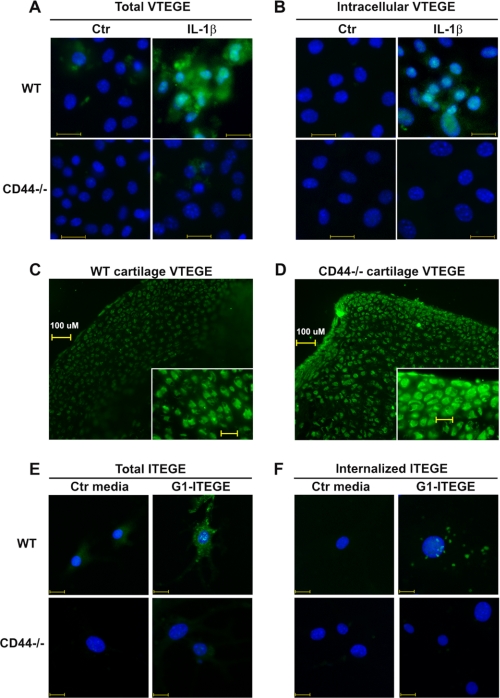
FIGURE 6.
Binding and internalization of endogenous G1-VTEGE or exogenous G1-ITEGE domains by wild type and CD44 null murine articular chondrocytes. Murine articular chondrocytes, grown on chamber slides, were incubated in the absence or presence of 10 ng/ml IL-1β under serum-free conditions as labeled. Panel A represents total G1-VTEGE, whereas panel B represents intracellular G1-VTEGE revealed after testicular hyaluronidase treatment to remove cell surface-associated G1-VTEGE. Wild type chondrocytes (WT) and CD44−/− cells are labeled as shown. Cells were fixed, permeabilized, and immunostained for G1-VTEGE, mounted in medium containing DAPI, and visualized by fluorescence microscopy. Shown are digital overlay images of green and blue fluorescence channels. In a separate study, explants cultures of wild type (panel C) or CD44−/− (panel D) femoral heads were incubated with 10 ng/ml IL-1β for 7 days. The tissue explants were then fixed, embedded, cryostat-sectioned, and immunostained for G1-VTEGE domains. In panels E and F, passaged murine articular chondrocytes, grown on chamber slides, were incubated with control media (Ctr) or G1-ITEGE-containing medium derived from bovine cartilage explants cultures. Cells were fixed and immunostained for G1-ITEGE, as the cells shown in panels A and B. Panel E represents total G1-ITEGE retained, whereas panel F represents intracellular G1-ITEGE, revealed after testicular hyaluronidase treatment to remove cell surface-associated G1-ITEGE. Bars in panels A, B, E, and F indicate 20 μm.

