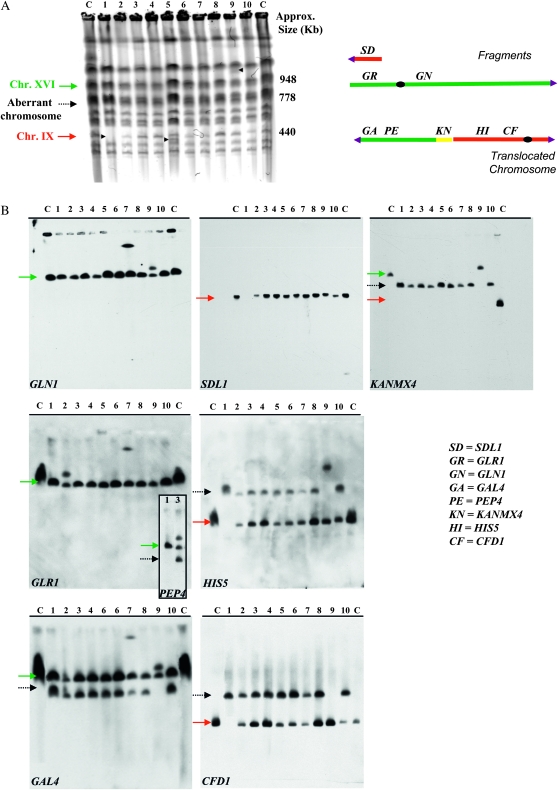
Figure 2.—
Chromosome analysis of the SUSU translocants. (A) Chromosome separation by CHEF. Arrowheads point to visible chromosomal polymorphisms in the SUSU1, SUSU5, and SUSU9 translocants. (B) Southern blot analyses. Blots were hybridized to the labeled GAL4, PEP4, GLR1, and GLN1 probes on chromosome XVI; SDL1, HIS5, and CFD1 probes are on chromosome IX; the KANMX4 probe was use to detect the aberrant chromosome. In all Southern blots, with the exception of that hybridized with KANMX4, lane C contains San1 diploid control strain DNA. In the membrane hybridized with KANMX4, the lane C contains San1-(DBP1/dbp1∷KANR) DNA; lane 12 contains San1-(GUT2/gut2∷KANR) DNA. Lanes 1–10 represent DNA derived from SUSU1 to SUSU10. The adjoining PEP4-probed membrane on the GLR1 panel shows the SUSU2 chromosomal pattern in comparison to the control strain San1. All the other translocants have the same pattern as in the GAL4 panel. Red arrow: chromosome IX; dashed arrow: translocant chromosome; green arrow: chromosome XVI.

