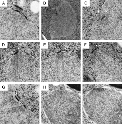
Figure 8.—
Electron microscopy analysis of SPBs in cells lacking Mps3p. (A) EM image from pom152Δ cells (SLJ4260) of duplicated side-by-side SPBs connected by a bridge. Asterisk indicates vesicle structures in close proximity to the SPB. (B–H) EM images from pom152Δ mps3Δ mutant cells (SLJ4259). (B and C) Low magnification (B) and high magnification (C) image of SPB present on a nuclear invagination. An arrowhead points to electron-dense material resembling a satellite, and an arrow marks a cytoplasmic microtubule emanating from the half-bridge region of the SPB. (D) An unduplicated SPB with a half-bridge that appears to have lost association with the nuclear envelope (arrowhead). (E and F) SPBs that contain electron-dense material resembling a satellite (E, arrowhead) or duplication plaque (F, arrowhead) at the distal tip of the half-bridge. (G) Duplicated side-by-side SPBs connected by a bridge. Asterisk indicates vesicle structures located in close proximity to the SPB. (H) Serial section images showing a bipolar spindle. The first SPB is apparent in H (left) while the second SPB is apparent in H (right). Bars, 100 nm.

