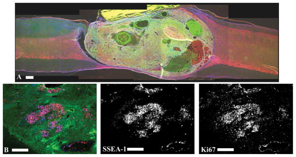
Figure 2.
Over-proliferation of transplanted ESNPCs. A subset of transplanted ESNPCs over-proliferated damaging uninjured spinal cord axons and cause a dramatic decrease in behavioral function. A) Over-proliferation was characterized by growth of ESNPCs that expanded from the ventral to the dorsal surface of the spinal cord. The image shows a sagittal section of a spinal cord with ESNPCs over-proliferation. The section was stained with Tuj1 (neurons, red), GFAP (astrocytes, blue) and GFP (ESNPCs, green). B) The proliferation marker Ki67 was used to visualize proliferating cells at 4 and 8 weeks after transplantation. The vast majority of Ki67 staining (blue) is co-localized with SSEA-1 (red) staining. The staining suggests that SSEA-1 positive mouse embryonic stem cells are contributing to the over-proliferation.

