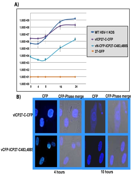
FIG 3 .
CFP–ICP27-C483,488S is confined to the nucleus during infection. (A) Vero cells were infected with HSV-1 KOS, 27-GFP, vICP27-C–CFP, and vN-CFP–ICP27-C483,488S at an MOI of 1. Experiments were performed in triplicate, and virus was harvested at 0, 4, 8, 16, and 24 h after infection. Plaque assays were performed in duplicate on Vero cells. (B) RSF were infected with vICP27-C–CFP or vN-CFP–ICP27-C483,488S at an MOI of 10 for 4 and 10 h after infection. CFP fluorescence was viewed with a Zeiss LSM 510 Meta confocal microscope at a magnification of ×63.

