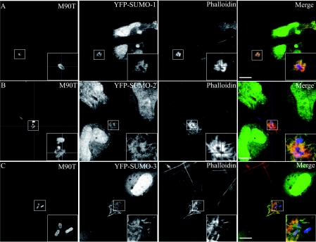
Figure 1. Distribution of galectin-3 and SUMO during the early stages of Shigella infection of epithelial cells.
HeLa cells were transfected with YFP (yellow fluorescent protein)–SUMO-1 (A), YFP–SUMO-2 (B) or YFP–SUMO-3 (C) proteins and then infected with S. flexneri M90T for 30 min at 37°C. Infected cells were next fixed and processed for labelling. M90T bacteria and galectin-3 were visualized by immunostaining with specific primary antibodies and then a Marina-conjugated or an Alexa Fluor®555-conjugated secondary antibody respectively. Scale bar, 10 μm.

