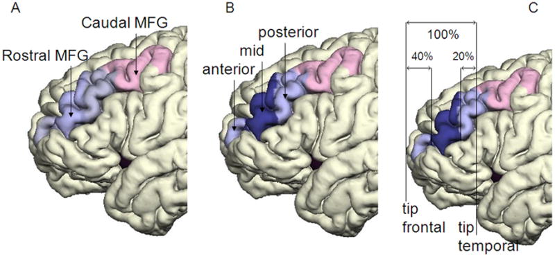
Fig. 2. Pial surface reconstruction representing the parcellated subregions of MFG.

Panel A shows caudal MFG (colored pink) and rostral MFG (light blue). Panel B shows the three subregions of the rostral MFG: the anterior rostral MFG, mid-rostral MFG (MR-MFG)(dark blue) and posterior rostral MFG. Panel C demonstrates the anterior and posterior boundaries of MR-MFG, adapted from Al-Hakim and colleagues (Al-Hakim, 2006).
