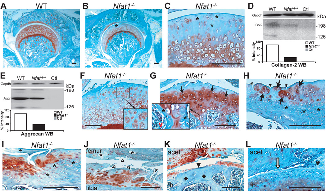
Figure 1.
Nfat1 deficiency causes OA-like changes in weight-bearing appendicular joints of adult mice. All photomicrographs are representative of at least 5 Nfat1−/− or WT mice per gender at each time point. Safranin-O and fast green staining and counterstained with hematoxylin. Scale bar = 200 µm. (A) A 2-month-old female WT mouse hip joint showing normal joint structure and articular surface with safranin-O staining (red) for proteoglycans throughout the entire articular cartilage. (B) At 2 months of age, a focal loss of safranin-O staining for proteoglycans is evident in the superficial/upper zone (*) of femoral head articular cartilage of a female Nfat1−/− mouse. Formation of abnormal periarticular cartilage is not evident at this stage. (C) Higher magnification of (B). (D) At 2 months, Western blot (WB) using a polyclonal antibody against human (cross-reacts with mouse) collagen-2 (Col2, Santa Cruz, sc-7764) with semi-quantitative measurements of band intensity reveals a loss of collagen-2 in female Nfat1−/− femoral heads. WB using Gapdh antibody (IMGENEX, IMG-3073) was used as a loading control. Ctl represents a negative control without using primary antibody. (E) At 2 months, a severe loss of aggrecan in female Nfat1−/− femoral heads was detected by WB using a polyclonal antibody against human (cross-reacts with mouse) aggrecan (Aggr, Santa Cruz, sc-25674) with semi-quantitative measurements of band intensity. (F) At 4 months, roughening of the articular surface, focal loss of proteoglycan staining, and early chondrocyte clustering (in magnified square) are seen in the upper-mid zones of articular cartilage of a female Nfat1−/− hip. (G) A 6-month Nfat1−/− hip joint shows roughening and discontinuity of articular surface, loss of safranin-O staining in the upper zone, chondrocyte clustering in the upper-mid zones (arrows), and increased safranin-O staining in mid-deep zones of femoral head articular cartilage. Endochondral ossification, including chondrocyte differentiation/hypertrophy, vascular invasion (red v), and new bone formation (*) on the surfaces of calcifying cartilage, is seen in subchondral bone (in magnified rectangle). (H) A 12-month Nfat1−/− shoulder (glenoid) shows chondrocyte clusters (arrows) with thickened subchondral bone (*) and vertical clefts in the superficial zone of articular cartilage (arrowheads). (I) A 15-month Nfat1−/− humeral head displays a fragment of cartilage (♦) derived from deteriorated articular cartilage due to a combination of horizontal and vertical fissures/clefts. Increased proteoglycan staining (red) is evident in the areas adjacent to the fissures. Thickened subchondral bone (*) is clearly seen. (J) At 15 months, articular cartilage destruction with surface fibrillation (△) and subchondral bone cysts (*) are seen in an Nfat1−/− knee joint. (K) An 18-month Nfat1−/− hip shows thinning or complete loss of articular cartilage (eburnation) (arrowhead) with exposure of thickened subchondral bone (♦). acet, acetabulum; fh, femoral head. (L) An 18-month Nfat1−/− hip shows narrowing of joint space (open arrow) and fibrous joint fusion (♥).

