Figure 3.
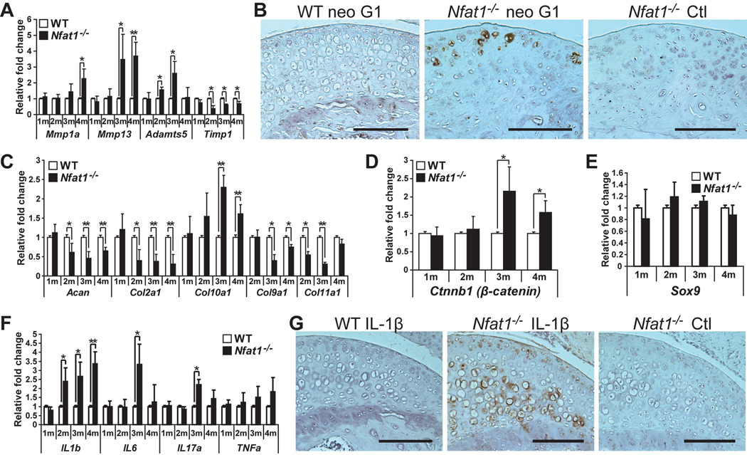
Loss of Nfat1 leads to an imbalance between catabolic and anabolic activities of articular chondrocytes favoring catabolism. (A) qPCR analyses of genes of interest at 1–4 months (1–4m) demonstrate up-regulated expression of Mmp1a, Mmp13, and Adamts5 and reduced expression of Timp1 in femoral head articular cartilage of female Nfat1−/− mice compared to age-matched female WT mice. n = 3 pooled RNA samples, each prepared from the articular cartilage of 6–8 femoral heads; * P < 0.05; ** P < 0.01 for all qPCR analyses in Figure 3. (B) Immunohistochemical staining using an antibody (1:50) against human (cross-reacts with mouse) aggrecan neo G1 (Affinity BioReagents, PA1-1746), which detects the degraded aggrecan product, shows that staining of degraded aggrecan (brown areas) was substantially more intense around cells in the upper zone of 3-month old female Nfat1−/− femoral head articular cartilage than age-matched WT mice. Nfat1−/− Ctl represents a negative control without using neo G1 primary antibody. Scale bar = 200 µm for (B) and (G). (C–F) qPCR analyses indicate temporal changes in expression levels of various genes of interest in Nfat1−/− articular cartilage at 1–4 months. n = 3 pooled RNA samples, each prepared from the articular cartilage of 6–8 femoral heads. (G) Immunohistochemical analyses using a polyclonal antibody (1:50) against rat (cross-reacts with mouse) IL-1β (Santa Cruz, sc-1252) show substantially more intense expression of IL-1β (brown areas) in femoral head articular cartilage of 3-month-old Nfat1−/− mice compared to age-matched WT mice. Nfat1−/− Ctl represents a negative control using both IL-1β antibody and IL-1β blocking peptide (1:100) (Santa Cruz, sc-1252P) to validate the specificity of the immune reaction in Nfat1−/− articular cartilage.

