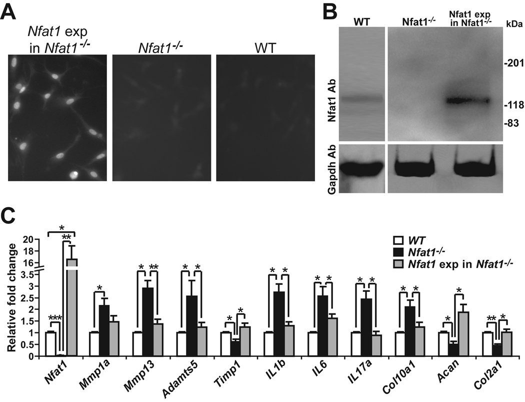
Figure 4.
Forced expression of Nfat1 rescues abnormal activities of 3-month-old Nfat1−/− articular chondrocytes. (A) Immunofluorescence using an antibody against hemagglutinin (HA) tags shows positive expression of Nfat1 protein in the nuclei of articular chondrocytes after lentiviral DNA transfection of constitutively active (CA) Nfat1 plasmid with HA tags from Addgene (Nfat1 exp in Nfat1−/−), but not in control Nfat1−/− or WT articular chondrocytes in which HA tags are not present. Cultured cells for immunostaining were fixed with cold methanol for 15 minutes and incubated with the anti-HA-fluorescein high affinity (3F10) antibody (Roche, Cat.1988506) at a concentration of 5µg/ml in PBS for 1 hour at 22°C. After washing three times with PBS, the cells were observed and photographed using a Nikon (ECLIPSE TE300) fluorescence microscope equipped with a Spot RTKE camera. (B) Western blots using Nfat1 antibody (Nfat1 Ab) confirm the effectiveness of forced expression of CA-Nfat1 protein in cultured articular chondrocytes. Western blots using Gapdh antibody are presented as loading controls. (C) qPCR analyses indicate the expression levels of Nfat1 and other genes of interest in cultured WT, Nfat1−/−, and Nfat1 exp in Nfat1−/− articular chondrocytes. n = 3 cultures. * P < 0.05; ** P < 0.01; *** P < 0.001.

