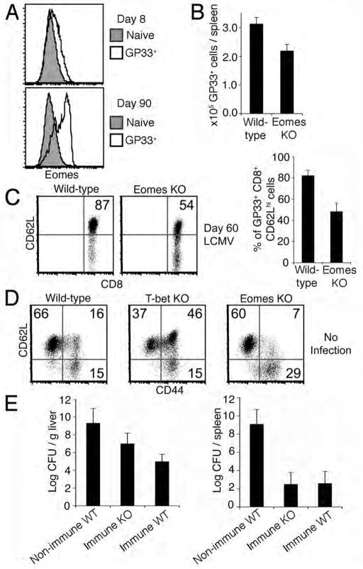
Figure 1. Diminished central-memory CD8+ T cell compartment in the absence of Eomes.
(A) Eomes expression in effector (8 days post LCMV infection, GP33+) and memory (90 days post LCMV infection, GP33+) CD8+ T cells relative to naïve (CD44lo GP33-) CD8+ T cells from spleens of C57BL/6 mice.
(B) Quantification of CD8+ GP33+T cells harvested from spleens of wild-type and Eomes KO mice 60 days after infection with LCMV. Data in (A) and (B) are representative of three independent experiments.
(C) GP33-specific central-memory CD8+ T cells in spleens of wild-type versus Eomes KO mice 60 days after infection with LCMV. Plots show CD8+CD44 +GP33 + cells. Numbers within plots refer to percentage of cells in the upper right quadrant. Graph shows mean +/− SEM from six mice of each genotype.
(D) Central-memory (CD62Lhi CD44hi), effector-memory (CD62LloCD44 hi) and naïve (CD62Lhi CD44lo) CD8+ T cell compartments in spleens of 6–12 month old naïve, non-infected wild-type, T-bet KO, or Eomes KO mice. Numbers within plots refer to percentage of cells in the respective quadrant. Data are representative of three independent experiments.
(E) Listeria monocytogenes-GP33 (LMgp33) burden in liver or spleen 3 days after infection with 5×10⁵ LMgp33 organisms in non-immune mice (non-immune WT), immunized (by LCMV infection 100 days prior) wild-type mice (immune WT), and immunized Eomes KO mice (immune KO) with three mice per group. Data is representative of two independent experiments.

