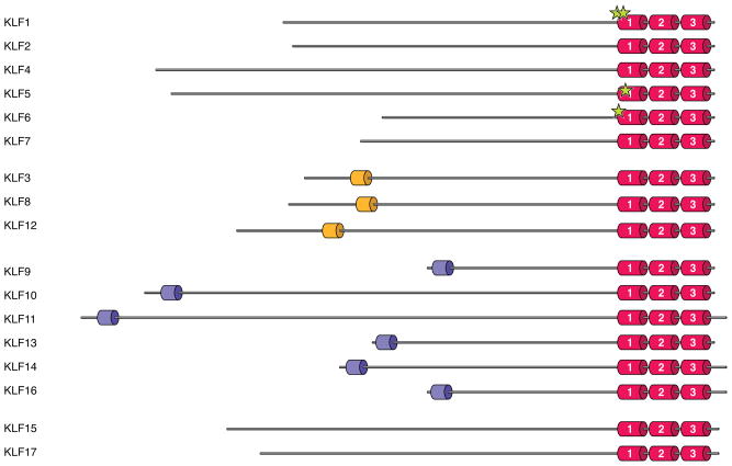
Figure 2. Protein structure of human KLF family members.
KLF proteins are grouped according to common structural and functional domains. KLFs are highly homologous in their carboxyl-terminal DNA-binding regions, which contain three C2H2 zinc finger motifs. The family members were grouped based on: (1) the ability to bind acetylases (KLFs 1, 2, 4, 5, 6, and 7); (2) the presence of a CtBP-binding site (KLFs 3, 8, and 12); or (3) the presence of a Sin3A-binding site (KLFs 9, 10, 11, 13, 14, and 16). Established sites of acetylation are marked by stars.

