
Figure 3. Localization of Klf4 and Klf5 in the mouse colon.
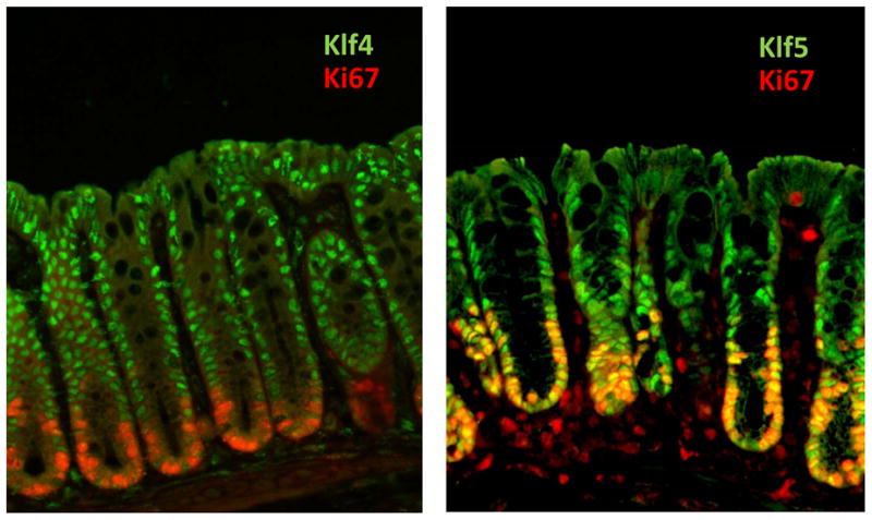
Immunofluorescence staining of Klf 4 or 5 (green) with the proliferation marker, Ki67 (red), was conducted on mouse colon. Klf4 (green) is present in the nuclei of terminally differentiated epithelial cells in the upper regions of colonic crypts. In contrast, Klf5 is localized to nuclei of proliferating epithelial cells at the base of the crypts. Ki67 highlights regions of active proliferation. Although Klf4 and Ki67 staining patterns do not overlap, Klf5 and Ki67 exhibit considerable co-localization, indicated by yellow staining.