
FIGURE 1.
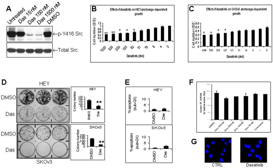
Dasatinib inhibits Src tyrosine phosphorylation, cell proliferation and colony formation of human ovarian cancer cells with minimal induction of apoptosis in vitro. (A), Effect of dasatinib (Das) on tyrosine phosphorylation of Src at tyrosine 416. HEY cells were treated with dasatinib or DMSO for 24 hrs and total protein was then extracted for Western blotting. (B), Effect of dasatinib on HEY growth. HEY cells were treated with dasatinib for 72 hrs. Crystal violet staining expressed as optical density (OD) was used to measure cell proliferation. *, P < 0.05 compared to untreated group. **, P < 0.01 compared to untreated group. (C), Effect of dasatinib on SKOv3 growth. SKOv3 cells were treated and evaluated as described in Fig. 1B. *, P < 0.05. (D), Effect of dasatinib on colony formation. HEY and SKOv3 cells were treated with dasatinib for 14 days. **, P < 0.01. (E), Effect of dasatinib on apoptosis in HEY and SKOv3 cells as determined by Sub-G1 fraction. HEY and SKOv3 cells were treated with dasatinib (150 nM for HEY, 300 nM for SKOv3) for 24 hr. (F, G), Effect of dasatinib on caspase activity and nuclear morphology. HEY cells were treated with dasatinib for 24 hr. Caspase 3/7 activity (F) was expressed as relative luminescence unit (RLU). DAPI was used to stain nuclei (G).