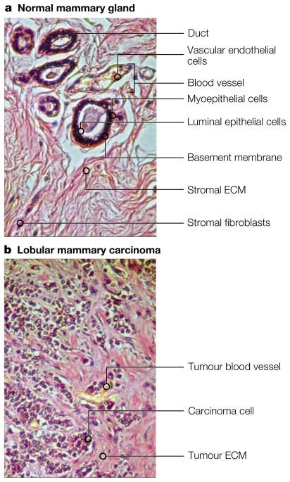
Figure 1. Normal versus malignant breast tumours.
a. The normal mammary gland shows a highly structured and segregated architecture. Ducts are formed by a double layer of cells: luminal epithelial cells surrounded by a layer of myoepithelial cells, enclosed by the basement membrane. Stromal fibroblasts secrete a collagenous extracellular matrix (ECM), and blood vessels are centrally located and well defined. b. Lobular breast carcinoma is less organized. Tumour angiogenesis produces poorly defined blood vessels, and carcinoma cells intermingle with all the stromal elements.

